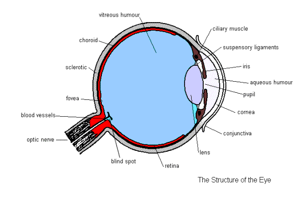
Eye Structure

- Aqueous humour
- a transparent watery fluid in the front part of the eye.
- Conjunctiva
- a thin, transparent membrane covering the inside of the eyelids and the front of the eye.
- Cornea
- the transparent bulge in front of the eye. Being transparent, it allows light rays to enter the eye. This is a continuation of the sclera. Iris- coloured part of the eye which surrounds the pupil a continuation of the choroid. It regulates how much light enters the eye.
- Vitreous humour
- transparent jelly which fills the large space behind the lens. It keeps the lens and the retina in position and also helps in the bending of light.
- Lens
- a transparent disc which focuses the light on the retina.
- Pupil
- the hole in the middle of the iris through which light passes.
- Ciliary muscle
- a ring of muscle which alters the thickness of the lens do rays can be focused on the retina. When it contracts the suspensory ligaments become slack and the lens thickens and when it relaxes the lens becomes thinner as the suspensory ligaments pull on the lens.
- Choroid layer
- the thin black middle layer containing the main arteries and of the eye. The black pigment in this layer prevents reflection of light within the eyeball.
- Sclera
- tough, white, outer layer which protects the delicate parts inside the eye.
- Suspensory ligaments
- hold the lens in position and alter its shape in conjunction with the ciliary muscle
- Fovea
- also known as the yellow spot, this area has a large concentration of light sensitive cells and so has the greatest visual ability.
- Retina
- the inner most layer of the wall of the eye, containing light sensitive cells.
- Optic nerve
- connects the eye to the brain.
- Blind spot
- the point at which the optic nerve leaves the eye. It is so called as there are no light sensitive cells present here.
- Blood vessels
- supplies the eye with food and oxygen.
